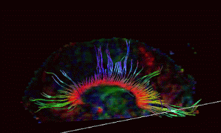
This series on Advances in Neuroimaging began with a quote from an article describing recent developments with Diffusion Tensor Imaging, or DTI. DTI is essentially a technique that allows us to see pathology in what are called fiber tracts within the white matter of the brain, even though individual axons are too small to be see without a microscope. If you have ever see the an insulation removed phone cable (as opposed to a telephone wire) you will see that inside the large cable, there are many small and colored
Smaller wires. This is analogous to the axon tracts within the white matter of the brain. Even though individual axons are microscopic, they tend to run in pathways with other axons, making a configuration that is large enough to image, with the right imaging method. That imaging method is DTI.
Within the brain, the water molecules tend to conglomerate and move, along the contours of the axon or fiber tracts. If you were to get a several pieces of string wet and then hold them vertically, you would probably see the water running down each strand. This is essentially what can be imaged in DTI. When diffuse axonal injury occurs in concentrated areas in the brain, even though we cannot see any of the individual axons that are damaged, we are able to see an interruption in the flow of water molecules along these fiber tracts. While DTI is certainly not showing all of the axonal damage throughout the brain, it is one more tool to show pathology, and in recent studies may be a strong indicator of those concussions which cause persisting and permanent deficits.


