Advances in Neuroimaging – Value in Diagnosis of Brain Injury

0 comments

Posted on 31st March 2008 by Gordon Johnson in Uncategorized

, , , , , ,

Last week’s blogs covered a series of articles about the advances in neuroimaging to assist in the diagnosis of subtle brain injury (otherwise called Mild Traumatic Brain Injury or concussion.) The key to looking back at those blogs, is the word “assist.” Imaging studies can only tell us what the structures of the brain look like. They cannot tell us how they got that way. They tell us very little about the function of the brain (although that may change dramatically with the continued development of fMRI.)

The first time a client of mine got an abnormal 3T MRI, I was so ecstatic, I thought my job had completely changed. It didn’t. The words “clinical correlation required” became an integral part of each case and frankly, it is a good thing it did. “Clinical correlation required” in essence means that did this person suffer a change in the way his brain was functioning, at a point in time consistent with the pathology that is being seen on the scan.

That is what being a brain injury lawyer is all about. Taking the technical findings of various subspecialist in the field of brain injury and putting them in front of a jury in a way that the jury can clearly see that the traumatic event, resulted in a change in this person, which is clearly related (correlated) to the brain damage that could be suffered in the accident. Without the real world picture of how this human being has been changed, with the line of demarcation of the accident, one can simply not make a diagnosis of brain injury.

I have been saying that same thing since I first wrote a web page on brain injury in 1996. Here is the words and the graphics I said at that time:

They are:

1.
Sufficient Biomechanical Force;

2.
One of the Four Acute Symptoms of the Rehab Congress’s definition, i.e.:

a) any period of loss of consciousness,
b) a change in mental state as a result of the accident,
c) amnesia, or
d) focal neurological deficits;

3. Neuropsychological Deficits; and

4.
A Changed Person.

Click here for those words I first wrote in 1996.

Now, we have more cases than we did in 1996 where the neuroimaging is abnormal. Yet, we still must show the same things: an accident with the potential to injure the brain, acute evidence that the brain was injured, deficits that can be determined in how a person functions and a CHANGED PERSON. Neuroimaging adds to the equation, but doesn’t eliminate any of the other issues. The only thing I would seriously change from what I said in 1996 is that there are other ways in addition to neuropsychological assessment, that deficits in ways in which the brain are working, can be identified.

Diffusion Tensor Imaging

0 comments

Posted on 29th March 2008 by Gordon Johnson in Uncategorized

, , , , ,


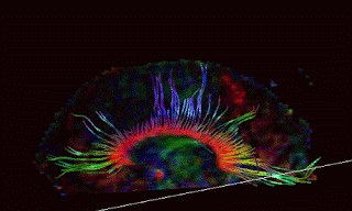
This series on Advances in Neuroimaging began with a quote from an article describing recent developments with Diffusion Tensor Imaging, or DTI. DTI is essentially a technique that allows us to see pathology in what are called fiber tracts within the white matter of the brain, even though individual axons are too small to be see without a microscope. If you have ever see the an insulation removed phone cable (as opposed to a telephone wire) you will see that inside the large cable, there are many small and colored
Smaller wires. This is analogous to the axon tracts within the white matter of the brain. Even though individual axons are microscopic, they tend to run in pathways with other axons, making a configuration that is large enough to image, with the right imaging method. That imaging method is DTI.

Within the brain, the water molecules tend to conglomerate and move, along the contours of the axon or fiber tracts. If you were to get a several pieces of string wet and then hold them vertically, you would probably see the water running down each strand. This is essentially what can be imaged in DTI. When diffuse axonal injury occurs in concentrated areas in the brain, even though we cannot see any of the individual axons that are damaged, we are able to see an interruption in the flow of water molecules along these fiber tracts. While DTI is certainly not showing all of the axonal damage throughout the brain, it is one more tool to show pathology, and in recent studies may be a strong indicator of those concussions which cause persisting and permanent deficits.