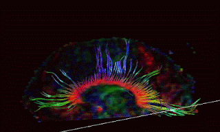
My topic for the rest of this week is advances in diagnosing brain injury thru improved neuroimaging. A recent study out of BYU, highlights some of the exciting changes that occurring, “New study shows brain changes from concussion
By Elaine Jarvik,
Deseret Morning News Published: Monday, March 17, 2008.
See http://deseretnews.com/dn/view/0,5143,695262379,00.htmlThe Deseret article begins:
“Even after a severe concussion, a brain can look normal and healthy on a traditional brain scan. But now a study co-authored by a Brigham Young University psychology professor, using a new kind of MRI technique, reveals brain changes that are subtle but significant.”
This article is talking about a technology called DTI imaging, but to fully understand the advances in neuroimaging, it is necessary to understand some basics about the science of neuroimaging and improvements in both the magnets and the software to interpret the raw information has changed.
The last three years have been an exciting time to be a brain injury lawyer because the implementation of 3 Tesla MRI scanners for clinical diagnosis of mild brain injury has resulted in an exponential increase in the number of abnormal scans for our clients. But increased field strength is part of the equation.
INCREASED FIELD STRENGTH
Tesla is the measurement of the strength of a magnet. 1.5 Tesla (1.5 T) is the current prevalent maximum field strength of MRI scanners found in US hospitals, with many facilities having scanners with weaker field strengths. While research facilities have been using considerably stronger field strengths than the 1.5 for at least five years, it wasn’t until mid 2004, that 3 T MRI scanners began to appear for clinical use. As I write this in March of 2008, there is likely a 3T MRI scanner at most major university medical centers, although many of these may still be restricted to research only applications.
One way to conceptualize the improvement in scanners is to compare such to similar improvements in the mega pixel capacity of a digital camera. An 8 mega pixel camera has roughly twice the resolution of a 4 mega pixel camera, and while the difference in MRI scanners don’t quite track a pure arithmetic improvement, the analogy holds quite nicely. After all, MRI scanners are essentially cameras, that use as the contrast agent, the vibrations of magnetized protons, instead of light.
My examination of a leading neuroradiologist, will a bit technical, will assist those who want to understand the details of these new advances:
My examination of a leading neuroradiologist in a recent case, may be helpful to understand the basic principles:
23 Q. My understanding is that MRI
24 imaging essentially uses an especially powerful
25 magnet with respect to 3-T to make the
1 molecules inside the brain resonate; is that
2 correct?
3 A. Correct.
4 Q. Explain what’s really going on
5 there.
6 A. What happens with an MRI
7 examination — for example, you mentioned
8 specifically 3-T. Well, the T stands for
9 Tesla. The more — the higher the Tesla
10 number, the more power the magnet. Which
11 really translates to your ability to see
12 smaller things.
13 So in many ways it’s analogous to
14 a microscope. If you have a higher powered
15 microscope you can see things better than you
16 can a lower powered microscope. An MRI
17 scanner is a higher powered. An MRI scanner
18 you can see things — many things you can
19 see better.
20 It’s not absolutely universal that
21 you see everything better, but for the most
22 part you see things much better on a higher
23 field strength magnet.
24 No matter what field strength
25 magnet you’re in, if I put you in an MRI
1 machine, basically what happens is that the
2 protons, which are part of the water
3 molecule, tend to line up with a magnetic
4 field.
5 So right now your water molecules
6 and your protons are just random in the
7 direction. They have a direction, and that
8 direction is random all over the place.
9 When I put you in an MRI machine,
10 they all line up. They all line up with a
11 magnetic field. And then what we do is we
12 give a radio frequency pulse. And it’s
13 basically very, very similar to an FM radio
14 wave. It’s almost the same energy as an FM
15 radio wave.
16 And basically what we do is we hit
17 your body with what’s called a radio
18 frequency pulse, which is really similar to
19 an FM radio wave. So it’s not dangerous.
20 There’s nothing bad about it. But what it
21 does do is it knocks those protons out of
22 that alignment.
23 And then as those protons come
24 back into alignment, they come back into
25 alignment at different rates, different speeds
1 based on the tissue, which is referred to as
2 a relaxation time.
3 So that the time it takes for
4 those protons to come back into alignment is
5 different for the skin, for the bone, for the
6 skull, for the cerebrospinal fluid. They all
7 have different rates.
8 The computer then assigns a gray
9 scale. So it’s kind of like paint by numbers.
10 If the relaxation rate has a certain number,
11 then it gets a certain color.
12 So basically, the computer does
13 something that’s completely analogous to paint
14 by numbers, and creates a picture out of
15 that.
16 And we do that with different
17 settings, depending on what we’re looking for.
18 And we can emphasize different tissues.
Increased field strength is only part of the breakthrough in neuroimaging. As more and more pathology is seen on these scans, neuroradiologists are realizing that what were considered to be insignificant findings on lower field scans, are of the pattern and nature most likely explained by traumatic forces, not disease processes or normal variants.
Tomorrow:
Dilated Perivascular Spaces in Identifying Mild Brain Injury


