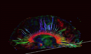
We began our series on “Advances in Neuroimaging” with yesterday’s blog on Increased Field Strength – the improvement from the 1.5 Tesla MRI’s to the 3 Tesla MRI’s. Today’s blog will discuss the evolving research that seemingly insignificant evidence of abnormalities on lower field strength scans, can illuminate evidence of traumatic injury. This is particularly true of what is technically called dilated perivascular spaces, also called Virchow Robbin Spaces. In essence, these are areas surrounding blood vessels in the brain, where myelin sheath or other brain tissue is missing, and thus show up as holes (white spots – also called UBO’s – unidentified bright objects) on the MRI scans. The technical term in an MRI report would be “areas of abnormal increased signal intensity.” Myelin sheath is the insulation type substance that protects the length of most multilevel axons in the brain.
Even though the 3T scans allow us to see these bright spots much clearer, this one term, “dilated perivascular spaces”, is still being used to describe lots of very different types of pathology. Again, excerpts from a recent deposition I conducted:
10 Q. We have the term called dilated
11 perivascular spaces that we’ve talked about.
12 Peri meaning?
13 A. Around the vessels. It’s the
14 spaces around blood vessels of the brain.
15 Q. And in a normal brain, what is in
16 those — why are there no spaces?
17 A. Well, there actually are spaces.
18 There’s spaces in everybody. It’s just that
19 they’re very, very small. And in some
20 patients you really see very few, or you
21 don’t really see hardly any, but they’re
22 there.
23 And then if they enlarge, because
24 you have lost substance in the brain around
25 them, then you refer to them as dilated
1 perivascular spaces, or enlarged spaces. And
2 what they fill in with is basically water;
3 cerebrospinal fluid is water.
4 Q. What type of brain matter is lost
5 in these, in the dilated perivascular
6 situation?
7 A. It’s basically a white matter
8 substance of the brain predominantly, because
9 that’s where you — that’s where these
10 perivascular spaces tend to be is mostly in
11 the white matter. So basically what you’ve
12 lost is some of the connecting fibers.
13 Q. Now, white matter is the axonal
14 part of the brain; is that fair?
15 A. Right. It’s the connecting fibers
16 of the brain. I made the analogy earlier of
17 the telephone systems. Telephone systems on
18 the surface of the brain, they’re basically
19 neurons. The connecting fibers are the
20 axons. And those connecting fibers are what
21 make up the white matter.
22 Q. And they call it white matter
23 because it’s white when you autopsy the
24 brain?
25 A. Depending on how you fix the
1 brain, yes.
2 Q. What is it that you’re seeing
3 that’s white? Is it the axons themselves or
4 the insulation around it?
5 A. It’s the insulation around them,
6 the myelins.
7 Q. And when we see dilated
8 perivascular spaces, are we seeing absence of
9 axons or absence of the insulation?
10 A. It could be both. We’re seeing an
11 absence of one or the other.
12 Q. Is the insulation considerably
13 larger in scale than the axons are?
14 A. Yes.
15 Q. Do you have any sense of the
16 magnitude of the difference?
17 A. An axon is on the order of about
18 50 microns. And then it has a mild sheath
19 around it. So that covering around that. So
20 the whole thing is really small.
21 Q. How small is a micron relative to
22 a millimeter?
23 A. It’s a thousandth of a millimeter.
24 Really tiny.
25 Q. So 50 microns would be 1/20th of a
1 millimeter?
2 A. Pretty small.
3 Q. Can you see axons in a human
4 macroscopically?
5 A. Only collections of them. Only
6 bundles of them. Large groups of them. You
7 can’t see 50 micron scales.
8 Q. And relative to the 3-Tessla MRI,
9 what is its resolution in terms of pathology
10 and the smallest pathology you see?
11 A. I think that depends on the type
12 of pathology. I would say in general you’re
13 in the 1 millimeter resolution range.
14 Depending on the pathology, you could go
15 smaller. Some pathology you might have to go
16 a little bigger. I feel very confident
17 calling 1 millimeter lesions.
18 Q. How many axons grouped together do
19 you think you would have to see to be able
20 to see it on the MRI?
21 A. Well, at the very least hundreds,
22 and probably thousands.
23 Q. Some are thicker than others?
24 A. Right.
For a more detailed explanation of the pathology of diffuse axonal injury, see http://subtlebraininjury.com/Neuropath
A dilated perivascular space on a 3T scan is no longer a vague bright dot but now has definition, measurable size and distinguishable shape. A neuroradiologist may be able to distinguish between such dilated spaces that can be caused by trauma, from other disease processes. .
25 Q. Is there weighting that goes into
1 your differential diagnosis when you look at
2 a dilated perivascular space in terms of more
3 likely trauma, more likely aging, more likely
4 microvascular? Is there — can you look at
5 the character in relation to the location of
6 perivascular spaces and shift a probability of
7 one diagnosis versus another?
10 THE WITNESS: Well, one of the
11 things that I’m looking at right now are
12 different types of perivascular spaces. And
13 we have a study that we’re conducting where
14 we’re looking at — I actually think there
15 are two types of perivascular spaces. There
16 are perivascular spaces that I would refer to
17 pathologic, and perivascular spaces that would
18 I would consider developmental.
19 So I’m going to eliminate the ones
20 that have — and what you’re asking me I’m
21 going to separate out the developmental ones.
22 The developmental ones, I think are
23 very round. They’re usually in the deep
24 basal ganglion region of the brain. They’re
25 very common. They can be extremely large.
1 They’ve been described in literature to be
2 well over a centimeter in size. Very big.
3 But I don’t think they ha
ve any clinical
4 significance at all.
5 Then there are dilated perivascular
6 spaces that I think are pathologic, meaning
7 that something caused them, whether that is
8 aging, whether that is a disease process,
9 whether that’s trauma.
10 I think that differentiating
11 between those requires you to look at a
12 variety of factors. And that is, does the
13 patient have any other disease condition?
14 What is the age of the patient? What are
15 the size of these perivascular spaces relative
16 to the age of the patient? Are they greater
17 than you anticipate for that patient’s age?
18 Is the location predominately in areas of the
19 brain where those particular disease processes
20 are most common? Do they fit together in that
21 way?
22 So my opinion is that we probably
23 will be able to over time improve our
24 differential diagnosis. I think we can put
25 it into two categories right now. Andhttp://www.blogger.com/img/gl.link.gif
then
1 I think beyond that it really requires
2 correlating it with clinical information, the
3 age, and the locatihttp://www.blogger.com/img/gl.link.gifon of the perivascular
4 spaces.
In summary, we have now covered improved field strength and dilated perivascular spaces. In tomorrow’s blog, we will address the need for tailored protocols in properly investigating Mild Brain Injury and the existence of Post Concussion Syndrome, aka, Subtle Brain Injury.


